Page 819 - My FlipBook
P. 819
PATHOLOGY OF THE DENTAL PULP.
By G. V. BLACK, M.D., D. D. S.
The dental pulp comprises the soft tissue that occupies the central
cavity of the crown of the tooth and the canals in the roots to the
apical foramen. It is thus divided into two portions—the coronal
portion or bulb, which occupies the crown-cavity, and the canal por-
tion, which occupies the roots or root-canals. Aside from this, the
coronal portion has a projection of its tissue under each of the cusps of
the tooth, as in the molars, which are called the horns of the pulp.
These horns are often quite long and slender, especially in young
teeth with long cusps. Generally, the form of the pulp corresponds
pretty closely to that of the tooth, except that it is every way more
slender.
The Tissue of the dental pulp is of the connective-tissue group, and
supports an abundant supply of blood-vessels and nerves. Its mass is
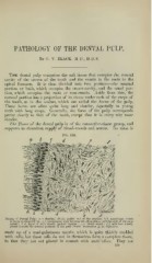
Margin of Dental Pulp: a, a, dentinal fibrils, pulled out of the dentine; b,b, membrana eboris
or layer of odontoblasts; r, c, transparent zone between the odontoblasts and the cells of the pulp
proper; d,(l, layer of cells closely packed together; r, f, blood-vessels; /,/, cells less closely
placed toward the central portions of the pulp (Wales' immersion ^ in. objective).
made up of a semi-gelatinous matrix, which is quite thickly studded
with cells, but these cells do not in themselves form a complete tissue,
in that they are not placed in contact with each other. They are
829