Page 820 - My FlipBook
P. 820
830 PATHOLOGY OF THE DENTAL PULP.
imbedded in the gelatinous matrix, always a little apart from each
other, even where most thickly set. The accompanying illustration (Fig.
440) gives a good idea of the pulp-tissue as seen with a high power in
thin sections stained with hsematoxylin. This is from the crown portion
of the pulp, and in this the cells are set in no particular position rela-
tively to each other, but seem to be placed as if by accident in every
conceivable position. In the root portion this is different : the cells
are there placed with their long axis parallel with the long axis of the
canal ; which arrangement gives the tissue quite a different appearance.
The Cells are generally spindle-shaped, with a delicate filament or
process extending from either end. The form, however, varies con-
siderably, especially in the coronal portion of the pulp. Some may
be seen so delicate and slender that they seem but little else than a
filament, while others are nearly round and much larger in their
central part. Again, we meet with many cells, especially in the
coronal portion, that have three and four filaments extending in as
many directions. In the normal pulp these filaments are very slender
and are lost in the gelatinous matrix. These, in all well-prepared sec-
tions, appear as minute threads in all parts of the tissue (as shown in
the illustration).
77?e Distribution of the Cells varies considerably in different portions
of the pulp. They are fewest in number in the central parts of the
coronal portion. All around the periphery of the pulp, just a little
inside the layer of odontoblasts, we find a zone that is much more
thickly studded with cells (r/). This is seen
Fig. 441.
in all parts of the pulp periphery. Between
this and the layer of odontoblasts there is a
narrow zone that is usually almost or quite
destitute of cells. In sections so prepared
as to show them this is found to be occupied
by a very fine plexus of nerves.
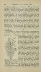
The Odovtoblasts form the periphery of
the pulp, and lie in contact with the dentine
(Fig. 440, 6). As seen with hematoxylin
staining, they seem to be flask-shaped cells
with a process extending into the dentine,
iiD^
the fibrils of Tomes, or the dentinal fibrils.
There is also a process extending from
the pulpal end of the cell which does not
\ take the stain and cannot be seen by this
mode of preparation. In Fig. 441, 1 have
shown these cells as they apj^ear in plain
unstained section, mounted in glycerin,
Odontoblast^ cliiuinR to i Fngnient with the one-sixteenth inch immersion
ot Iniperlecih clt\ eloped lieiitme.
llie ti^'iiie ^^ is pii kd ^^^ i\ in objective. The cells are shown just as
niuuiiiiiig tlitr ^tr^;tlull llit; uells
are drawn just as ihey lay distorted they happened to lie, without correcting
in the mounting, hut a good idea is
given of their true form (glycerin any of the distoi tion caused by the mount-
mounting, y'gth inch obj.).
ing. In this section the odontoblasts seem
to have been pulled oif from the tisssue of the pulp in pressing down
the cover-glass, and the fibrils are evidently somewhat stretched out of