Page 632 - My FlipBook
P. 632
642 DENTAL EMBRYOLOGY AND HISTOLOGY
note tlie difference in the two tnnies at the free margin of the enamel
organ. In yonnger specimens there is no apparent difference between
the two, but it must now be remembered that calcification has com-
menced at the apex of the tooth, and that material changes will now
occur in the inner tunic ; these have been noticed. The character of the
enamel organ as such is rapidly changing ; it has served its purpose,
and from now on, upon the apex of the papillae, it will disappear, and
this change will gradually proceed down the sides of the papillae until
the typal demands of the enamel cap are reached.
There is, however, a marked difference between the inner and the
outer tunic at this stage. The inner tunic gives evidences of rapid cell-
proliferation and consists of many
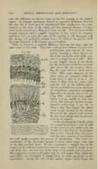
Fig. 366.
nuclei, forming a thick layer ; on
the other hand, the outer tunic con-
sists of a single layer of cells. This
is very happily shown in the figure
by the shrinkage which has occurred
just above the free margin, allow-
ing the outer tunic to stand out in
relief. This same character of the
outer tunic, as compared with the
inner tunic, is seen at circle c. At
circle d (Fig. 366) the outer tunic
and the cells of the stellate reticu-
lum have settled down upon the layer
of ameloblasts, sometimes arranging
themselves in whorls, seen at 7vh. ep.
(Fig. 358). Just what their signifi-
cation is I am unable to state pos-
itively, but from my studies in com-
parative embryology I am led to
believe that they supply the places
made by the increase in the circum-
ference of the enamel, and account
dn
for the short prisms seen in ground-
sections of enamel.
In the development of teeth, Avhere
the enamel is to form a coat of mail
Vertical Section tliroiifch .Spex of Central In-
cisor 10 cm. Porcine Embryo (X 500) : c. c/., con- on the crown of the tooth—viz. the
nective tissue of follicular wall ; /V, flat layer
of stratum intermedium; o, ameloblasts : Tp, Carnivora—the line of ameloblasts
Tomes processes into space; '/, enamel; dentirie; o, odontoblasts; dp, dental papilla.
sent the same number of ameloblasts
that will finally complete the process of calcification. The outer cir-
cumference of the developed enamel is many times larger than that
of the first calcified layer. If this represented a straight line, as the
enamel on the rodent's tooth does, then the space would be made at one
end of the line ; but here it is in the form of the greater part of a circle.
The expansion occurs at all parts, and the cell-supply from which the
amelol)lasts are developed is found lying in close proximity to the
ameloblasts. Along the side of the enamel orsran which forms the