Page 570 - My FlipBook
P. 570
580 DENTAL EMBRYOLOGY AND HISTOLOGY.
From the above quotations I think I am justified in saying that
all agree regarding the manner and method of intracartilaginous and
subperiosteal bone-formation, and intramembranous also, in so far as
it refers to the parietal Umes ; but, judging from the paucity of illus-
trations and literature treating directly upon the formation of the
remainder of the group not preformed in cartilage, I conclude that
there has been less investigation of this than of kindred subjects.
From my own studies in bone-formation, I am convinced that we
need all the classifications made by previous writers, and that still
another is essential to a clear understanding of ossified products, I
shall therefore make a fourth class, which I term intcj:stiti(d. In our
future study of bone, then, we shall have to consider four ways in which
it may be developed : I. In the substance of the embryonal connective
tissue of such bones as are not preformed in cartilage ; found in maxil-
lary ossification : interditicd (my own classification). II. In pre-existing
membranes, as in the skull-caj) : intramembranous. III. Underneath
the periosteum, as in the cortical portion of long bones : subperiosfeal.
IV. In pre-existing cartilage, as in the head of the fenuir : intracarti-
laginous.
I. Interstitial Formation of Bone.—I introduce this term for the ear-
liest development of bone, believing that it is needed. The term intra-
membranous, while applying to the manner of development in the
parietal and some others of the flat bones, does not apply to the
maxillae and bones of that class.
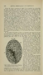
Fig. 320.
I am more convinced of the ne-
cessity of such a division of the
intramembranous group than of
the need of making a distinction
between the latter and subperios-
teal bone-formation. At best, in-
tramembranous ossification occu-
pies only a transitory stage, and
gradually passes into subperiosteal
by the ditferentiation of the peri-
osteum ; and I think a careful con-
sideration of the subject will so
convince the reader.
In the microscopic investigation
of early embryonic life (pig 2|- cm.,
and human 2 mo. ; see cut) cells are
seen grou])ed together here and
there in the central portions of
both the inferior and the superior
maxillfe, which have taken the
stain in such a manner as to call
Inferior Maxilla rortiiic iMiibryo {2]4 cm. X
210): o, osteoblasts t;'"oui)e(l together, siirrouiid- attention to their apjicarance in
;
ed by embryonal connective tissue, cl.
other words, they have been differ-
entiated by the staining process. ITnder low ))()wers thev do not difler
in f )rm iVoin the suridunding cells, but under high amplification they
show no processes as do the ordinary connective-tissue cells even at