Page 567 - My FlipBook
P. 567
OSSIFICATION. 577
composed of material unsusceptible of any change except that of disin-
tegration. The mollusk may add to its shell internally, but it nuist
necessarily be at the expense of the size of its own body. And so it is
with the osteoblasts: they are arranged in close proximity—so close
that the first secreted calcospherule jt)ins that of a neighboring sj)he-
rule, and by this juxtaposition and coalescence solid bone is formed.
Fracture of the shell may also be repaired by the same cells .which
in the first place secreted the shell.
The area of tissue supplied by a capillary vessel at the beginning of
the process of calcification marks the limit of the Haversian system.
The osteoblasts are arranged around the
Fig. 317.
outer portion of this area, antl the first-
formed layer of calcospherules consti-
tutes the periphery of the Haversian
system ; the next-formed layer of sphe-
rules lies inside the first-formed layer,
thereby lessening the space occupied by
the capillary vessels ; the third layer is
still inside the second ; and so on centrip-
etally, until the several layers almost
entirely fill the space (Fig. 316). The
remaining space is occupied by the vas-
cular antl lymphatic system, and no less
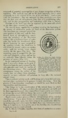
an authority than »Schaefer claims the Section of a Haversian Canal, showinR
its contents (highly magnified) : n, small
presence of nerves (Fig. 317). arterial capillary vessel ; v, large ven-
ous capillary; »., pale nerve-flbi'es cut
In the centripetal manner of develo})-
acioss; /, cleft-like lymphatic vessel:
ment I see a wise design on the part of one of the cells forming its wall com-
municates by fine branches with the
Nature to limit the space occupied by the branches of a bone-corpuscle. The sub-
stance in which the vessels run is con-
calcospherules and mark the outline of nective tissue with ramified cells ; its
finely granular appearance is probably
the Haversian system. This centrijietal
due to the cross-section of fine fibrils.
arrangement lessens the calil)re of the The caiiMl is surrounded by several con-
centric lamellie.
vessels, but yet allows them abundant
capacity to carry sufficient cell-pabulum to keep alive the enclosed
organic tissue.
Thus is cellular activity made self-limiting and a beautiful and sym-
metrical object conformed to its purpose brought into being. Were the
j)rocess of bone-formation centrifugal, we should be more likely to find
abnormalities and distortions.
This brings us to the consideration of the several forms in which
bone is developed. We have seen how the calcospherules are built and
bv their aggregation made into compact bony tissues, and it now remains
to discuss the several different forms they assume under the government
of pre-existing tissues which modify their arrangement.
Nearly every author gives a different interpretation of the existing
classifications. Upon tjiose known as intracartilaginous and subperi-
osteal they generally agree, but there seems to be considerable difficulty
in harmonizing their views upon the third class—viz. intramembranous.
This, as I shall try to show, grows out of the fact that this classification
is made to cover too nuich ground.
Dr. T. Mitchel Prudden describes this form of ossification as occur-
VoL. I.—37