Page 200 - My FlipBook
P. 200
210 ANATOMY.
rior bellies of the digastric muscle. It is covered externally by the
superficial layer of the submaxillary fascia, the platysma myoides mus-
cle, and the skin ; internally by its deep fascia, which separates it from
the mylo-hyoid, hyo-glossus, and stylo-glossus muscles. The gland
extends backward to the posterior border of the mylo-hyoid muscle,
where it sometimes passes around its border to the upper surface, and
is separated, at the posterior part, from the parotid gland by the stylo-
hyoid ligament.
The Hubmaxillari/ Dad (Wharton's), through which the secretion of
the above gland passes to the mouth, is about two inches in length,
and its coats are not so thick as those of the parotid duct. It com-
mences by the union of the ducts originating in the different lobules near
the posterior surface of the gland, and with some of the tissue of the
gland winds around the posterior border of the mylo-hyoid muscle. It
then passes forward and inward over the muscle and beneath the hyo-
glossus and the sublingual gland, terminating in a narrow opening
through a soft papilla at the side of the fraiuum linguae, near the duct
on the opposite side. Occasionally isolated lobules of gland tissue are
found along the duct.
The arteries which supply the gland are branches of the facial and
lingual. The veins belong to the facial and lingual.
Its nerve-supply is derived from the submaxillary ganglion, which
obtains its motor filaments from the chorda tympani, and its sensory from
the lingual branch of the inferior
maxillary—sometimes, though
Fig. 103. sel-
dom, from the mylo-hyoid, a branch
of the inferior dental ; the sympa-
thetic nerve branches from those
accompanying the arteries in this
vicinity.
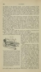
The Sublingual or Gland of Bar-
fhoiin (Fig. 103) is so named from
its position under the tongue. It is
smaller than the submaxillary gland,
and secretes mucus only. The lobules
View of the Right Submaxillary and Sublingual of jthis gland are not, as in the parotid
Cjlands, from the inside, i'art of the ritrlit
side of the jaw, divided from the left at the and submaxillary, united into one
symphysis, remilins; the tongue and its nius- with a single duct leading from
cle.s have been removed, and the mucous it,
membrane of the rifjht side has been dis- but are divided into several smaller
sected off aTid liHoked upward, so as to expose'
the sublingual glands; .v/», the larger super- glands, each having an independent
ficial part of the submaxillary gland;.;", the
facial artery passing through it; .vj?*',' deep duct. They are arranged in a nar-
portion prolonged on the inner side of the row, oblong form situated beneath
mylo-hyoid niuse'e, m/i ; xl is pbiced below
the antnrior large part of the sublingual the mucous membrane of the mouth,
gland, wiih the duct of f^artbolin partly
shown x/\ placed above the hinder small forming a ridge in the alveolo-lingual
;
end of the gland, ii dic;ites
;
(/, the papilla, at which the duct of Wharton front of the tongue near the franum,
opens in front behind tlie incisor teeth; the commencement of the duct ; A, the hyoid and in close proximity to the gland
bone ; n, the gustatory nerve ; close to it is
the submaxillary ganglion. of the opposite side ; it extends
backward and outward about one
and a half inches to near the iirst molar tooth. The inner surface of