Page 202 - My FlipBook
P. 202
212 ANATOMY.
a capsule and divided into two unequal portions, a superior or larger and
an inferior or smaller, with a thin layer of fascia between them.
The Siqjerior Portion, which comprises the greater part of the gland,
is firmer in structure than the inferior—made so by the larger size of
its lobules and their greater
^'
compactness of arrangement.
It is placed within the lach-
rymal fossa.
The Inferior Portion (glan-
dula lachrymalis inferior, Ro-
senmiiller) is smaller than the
superior. Its lobules are mi-
nute in size and loosely col-
lected together, which gives it
a softer appearance than the
superior portion. It is situ-
ated in the subconjunctival
connective tissue, just back
of the lateral portion of the
upper eyelid.
Harder's Gland, found in
most mammals, is situated at
the inner angle of the eye.
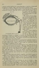
The Lachrymal Apparatus, right side. skeop, and
j^^ ^j^^ ^^^ pig,
according to Wendt, it is similar in structure and function to the lach-
rymal gland, while in the musk-rat and guinea-pig it is like the seba-
ceous glands of the body. This gland is found in man and in the ape
in a rudimentary state (Gracomini).
The Canmcula Lachrymalis is a small pinkish-red body situated at
the inner angle of the eye—the rudiment of the nictitating membrane
of birds, which forms a kind of third eyelid for protection, without
obstructing the functions of the organ.
The Buds of the Lachrymal Gland leading to the surface of the eye
are from ten to fourteen in number. They pass obliquely downward
beneath the mucous membrane, diverging slightly as they do so, and
finally open into the outer third of the superior palpebral sinus on a
line with each other. A few of the lobules composing the inferior
portion of the gland have independent ducts which open separately,
while others join the ducts coming from the superior or main portion
of the gland.
Arteries.—The lachrymal gland is supplied with blood through the
medium of the lachrymal artery, which is a branch of the ophthalmic.
Nerves.—The lachrymal gland receives its nerve-supply from the
lachrymal nerve, which is a branch of the ophthalmic or first division
of the fifth cranial nerve.
Tlie Lachrymal Canals, or Canaliculi, are four in number, a supe-
rior and an inferior for each eye. They have their origin in small
openings, the puncta lachrymalia, situated in the centre of a teat-like
elevation at the inner edge of each eyelid near the inner angle of the
eye the lachrymal papilla. These canals terminate separately by open-