Page 195 - My FlipBook
P. 195
—
'
AREOLAR TISSUE, TENDOJVS, AND MUSCLES. 205
shrunken remains of the mucous cells, consequent on exhaustion ; and
Klein is of the opinion that such is in reality the case ; for, arguing by
analogy, he finds that excessive stimulation results in structural changes
Avhich have already been noted, and accompanied by watery secretion
/. e. the cells have evidently discharged all their nuicin, and have col-
lapsed and become both morphologically and physiologically like those
of the true salivary glands. He also states that in the submaxillary
gland of young animals all gradations are met with from small alveoli
with small lumen lined only with small granular cells, and alveoli some-
what larger and lined either partly with mucous cells, partly with gran-
ular cells, or altogether with muccnis cells, to which are applied from
place to place groups of granular cells."
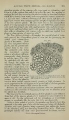
The True Salkanj Glandn.—Examples: the parotid gland of mam-
mals, parts of the submaxillary gland of man and the guinea-pig, the
orbital and submaxillary
Fig. 100.
glands of the rabbit. These
are also compound tubnlar
glands, and the general ana-
tomical form is the same.
The epithelial cells lining
the convoluted secreting
tubes or alveoli are different,
consecpiently their function
is not the same (Fig. 100).
Its epithelial cells are cubi-
cal, though the angles are
somewhat rounded ; they are
placed in a simple layer, con-
tain a spherical nucleus placed
near the basement-membrane,
and are united with those of
the cell proper, forming to-
Section of part of tlie Uunian Submaxillary filaiul. To the
gether irregular and small right ol the tigiiie is a group of luucous alveoli, to the
left a group of serous alveoli.
meshes. When the gland is
inactive these meshes contain a small quantity of fluid substance. In
osmic preparations the cell appears to be packed full of distinct granules
of an albuminous nature which obscure the nuclei.
Between the cells and basement-membrane there are quantities of
embryonal cells (crescents of Gianuzzi), though not so abundant as in
the glands last described.
The lumen in the convoluted portion of these glands is quite different
from that of the mucous glands is doubted by some whether it is
; it
open at all. In the embryonal cell the protoplasm or intercellular
cement so completely fills the tube that it is not discernible.
"After a short period of activity the granules are found to have dis-
appeared in the outer part of the cell, the inner part being still distinctly
granular, and some granules, being apparently free within the lumen
of the alveolus [tube], now becoming distinct (Fig. 101). With more
prolonged activity the clear outer part increases in extent, and ihe
' Coles's Microscopical Science.
'
AREOLAR TISSUE, TENDOJVS, AND MUSCLES. 205
shrunken remains of the mucous cells, consequent on exhaustion ; and
Klein is of the opinion that such is in reality the case ; for, arguing by
analogy, he finds that excessive stimulation results in structural changes
Avhich have already been noted, and accompanied by watery secretion
/. e. the cells have evidently discharged all their nuicin, and have col-
lapsed and become both morphologically and physiologically like those
of the true salivary glands. He also states that in the submaxillary
gland of young animals all gradations are met with from small alveoli
with small lumen lined only with small granular cells, and alveoli some-
what larger and lined either partly with mucous cells, partly with gran-
ular cells, or altogether with muccnis cells, to which are applied from
place to place groups of granular cells."
The True Salkanj Glandn.—Examples: the parotid gland of mam-
mals, parts of the submaxillary gland of man and the guinea-pig, the
orbital and submaxillary
Fig. 100.
glands of the rabbit. These
are also compound tubnlar
glands, and the general ana-
tomical form is the same.
The epithelial cells lining
the convoluted secreting
tubes or alveoli are different,
consecpiently their function
is not the same (Fig. 100).
Its epithelial cells are cubi-
cal, though the angles are
somewhat rounded ; they are
placed in a simple layer, con-
tain a spherical nucleus placed
near the basement-membrane,
and are united with those of
the cell proper, forming to-
Section of part of tlie Uunian Submaxillary filaiul. To the
gether irregular and small right ol the tigiiie is a group of luucous alveoli, to the
left a group of serous alveoli.
meshes. When the gland is
inactive these meshes contain a small quantity of fluid substance. In
osmic preparations the cell appears to be packed full of distinct granules
of an albuminous nature which obscure the nuclei.
Between the cells and basement-membrane there are quantities of
embryonal cells (crescents of Gianuzzi), though not so abundant as in
the glands last described.
The lumen in the convoluted portion of these glands is quite different
from that of the mucous glands is doubted by some whether it is
; it
open at all. In the embryonal cell the protoplasm or intercellular
cement so completely fills the tube that it is not discernible.
"After a short period of activity the granules are found to have dis-
appeared in the outer part of the cell, the inner part being still distinctly
granular, and some granules, being apparently free within the lumen
of the alveolus [tube], now becoming distinct (Fig. 101). With more
prolonged activity the clear outer part increases in extent, and ihe
' Coles's Microscopical Science.