Page 194 - My FlipBook
P. 194
204 ANATOMY. ;
sidered identical with them, having either migrated directly from the
blood-capillaries or from the lymphoid tissue surrounding the glands.
The ninnber of mucous corpuscles depends on the glands which produce
the mucus ; they vary according to locality.
III. Compound Tubular Salivary Glands.—These, generally
speaking, are six in number, three on each side, named sublingual, sub-
maxillary, and parotid.
Tlie minute anatomy and the physiological action of the salivary
glands prompted Lavdowsky to classify them into three groups: 1.
Mucous glands ; 2. True salivary glands ; 3. Muco-salivary glands.
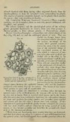
3Iucous Glands,—Examples : submaxillary and orbital glands of the
dog (Fig. 99) and cat and the sublingual glands of man. They are
similar to, though larger tlian,
Fig. 99.
the compound tubular glands of
the mucous membrane of the
mouth ; their general construc-
tion is the same, with the excep-
tion of the epithelial lining of
the convoluted portion of the
j^^ ^ ^ tube, which is composed of mu-
cous cells—the same kind of epi-
thelial cells as described in the
mucous membrane—and the pari-
etal cells, or crescents of Gianuzzi.
:
These cells are somewhat similar
to the embryonal cell already de-
scribed ; they stain deeper than
the surroundiny; tissue with hoem-
otoxylin and eosin, thus showing
their protoplastic condition ; they
are granular in ajipearance, and
Submaxillary Gland of the Dog: rt, mucous cells; ft, form somihinnr mnsSPS WlUKHlL
Avithoilt
^^^^^^ ^LUlllUliai
protoplasm cells; c, demilune of (iiaiuizzi; ,/, llUlbstiJi
transverse section of an excretory duct, with its definite membrane, but witll pi'O-
peculiur columnar epithelial cells. . ^ . ,-, , n , • , ,> • i
jections that fit into the uTegular-
sliaped spaces between the epithelial mucous cells and the basement-
membrane with which they are in contact. The lumen of the convo-
luted portion is o])en and of considerable size.
When tliese glands are stimulated to secretion, either through the
natural source or by artificial means, the mucous cells at first increase
in size, and a viscous fluid is secreted which passes out by the ducts
after the action has been continued a short time, the cell-nucleus changes
its shape and position, becoming smaller and granular, more rounded
and central, and the cell takes a deeper stain with carmine. If this
stimulation be ])rolonged until the glands become exhausted, the mucous
cells lo.'^e their identity, and are either lost by being carried oif in the
mucus, or they become granular and look like the parietal cells found
next to the basement-membrane.
" Heidenhain and Lavdowsky have asserted that they are destroyed,
and that their ])laces are taken by a process of new cell-formation from
the parietal areas ; but Ewald regards these smaller granular cells as the