Page 152 - My FlipBook
P. 152
162 ANATOMY.
having to pass from one extremity of the muscle to the other. But a
long muscle may be composed of a number of short fasciculi attached
obliquely to the sides of its tendon, which may advance upon its surface
or into its fleshy parts. Many short tiisciculi, thus connected, produce
by their combined operation a more powerful effect than a few fasciculi
extending the entire length of a muscle. The latter arrangement, how-
ever, gives greater extent of motion.
The Fibres composing the Fasciculi are cylindrical or prismatic in
form. Their size is generally uniform, being in the muscles of the
trunk and limbs from yjo^th to ^^th of an inch in diameter. It is
less in those of the head, especially in the face, where they range from
th to yyo-th of an inch.
2 4^)
The general length of the fibres does riot exceed an inch and a half.
In long fasciculi, therefore, they do not extend from the tendon of one
extremity to that of the other, but end in a rounded p(jint invested by
sarcolemma adhering to approximate fibres.
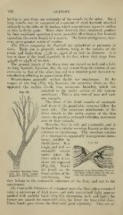
Muscle-fibres generally neither divide nor anastomose. In the
tongue of the frog (Fig. 83), however, the muscular fibres as they
approach the surface divide into numerous branches, which are
attached to the under surface of the mucous
Fig. 83.
membrane. This is also true of man and vari-
ous animals.
The fibres of the fticial muscles of mammals
and those of the panniculus carnosus follow the
same rule. The numerous attachments of the
latter muscle to the under surface of the skin
causes the peculiar external twitching movement
seen in these animals.
Muscular filjre is soft and contractile, and is
enclosed in a tubular envelope known as the sar-
colemma or myolemma. This envelope consists
of a transparent, apparently homogeneous mem-
brane, similar to
^^''- ^'^
elastic tissue. It is
tough, and will oc-
casionally remain
entire when the
fibres which it en-
closes are ruptured
(Fig. 84). Nuclei
are found on the
A Branched Muscular Fibre Fragments of an Elementary Fibre of
from the frog's tongue inner surface of the the Skate, held together by the un-
(magnified :!•">(( diameters). torii but twisted sarcolemma.
sarcolennna, but
they belong to the ccmtractile substance of the fibre, and not to the
sarcolemma.
The (bnfracti/e Suhsf(nicc of voluntary muscular fi1)re, when examined
under a microscope of high ])ower and A\ith transmitted light, appears
marked with parallel bands (Fig 8.)), alternating dark and light ; the
former are named the contractile discs, the latter the interstitial discs.
These bands pass across the fibx'e with great regularity. They are of