Page 154 - My FlipBook
P. 154
164 ANATOMY.
Blood-vessels in muscular tissue are extremely numerous. These earrv
the material for the nourishment of the tissues and for the chemico-vilal
changes which take place within them. When these vessels are filled
with coloring matter, the fleshy part of the muscles
Fig. Sfi
supplied by them is in strong contrast with the tendons.
Arteries, accompanied by veins, enter the muscle at
various points, divide into branches, pass among the
fasciculi, and break up more and more as they extend
into the finer divisions of the muscle. Finally, they
penetrate the smallest fasciculi and terminate in capil-
lary vessels which run between the fibres. They are
supported by the subdivisions of the perimysium, and
supply it with capillaries. The diameter of these is
extremely small, and they form a fine network among
the fibres.
Lymphatics.—It is not known that there are any
lymphatic vessels in the voluntary muscular tissue, but
they are found in great abundance in the connective
tissue of its sheaths and tendons. They have their
commencement in connective tissue, and their office is
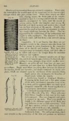
Muscular Fibre of a to collect and convey the lymph from the muscular
IManiinal, exaiiiiiied
fresh in serum, high- substance and tendons.
ly magnified, the sur-
face of the fibre be- The. Nerves of the voluntary muscles are of large
ing accurately focus-
ed. The nuclei are size, and their branches pass between the fasciculi, often
seen on the flat at uniting to form plexuses, from which smaller nerve-
the surface of the
fibre, and in profile filauieuts are givcu oiF and form finer plexuses, each
containing not more than two or three dark-bordered
nerve-fibres. Single nerve-fibres pass from these between the fibres of
the muscles, divide into branches, and finally terminate in motor end-
plates, which are situated upon the sarcolemna of the muscular fibres.
Small nerves also accompany the branches of
Fio. 87. blood-vessels within the muscle.
Involuntary Smooth or Unstriped Muscle.—
Excepting in the heart and a few other organs
of the body, involuntary muscular tissue is un-
^ striated, and its ap]:>arent fibres are made uj) of
elongated contractile cells bound together by a
homogeneous intercellular substance.
Unstriated muscular tissue is composed of con-
These cells may
tractile fibre-cells (Fig. 87).
form fibrous bundles or they may be less regu-
larly arranged. They are elongated, and usual-
ly pointed at the ends. They vary greatly in
length in the dififerent organs of the body, and
may bifurcate at one or both extremities. Each
cell has a nucleus, which is either oval or rod-
shaped, and situated, as a rule, centrally.
Involuntary Muscular Fibre-
cells from Human Arteries. Involuntary nuiscle fibre-cells are spindle or
fusifi)rm in shape. Tlie wall or envelope, whicli
may wrinkle on the contraction of the fibre and produce an indistinct