Page 90 - My FlipBook
P. 90
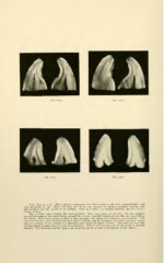
Figs. 44-a. b. c. d. These figures represent teeth that came to me split longitudinally and
very perfectly in line. They had been cut off for the purpose of placing artificial crowns, and
did not show the full length of the crowns. Figs. A, B and c are central incisors, and Fig. D is it
lateral incisor.
The surfaces were ground flat and polished ; they were then set up with the cut surfaces
toward the camera, the tooth being opened like a book, one-half laying on one side and one-half on
the other. They were photographed in this position with about six diameters enlargement. The
material did not make brilliant pictures, but it will be seen by scanning the labial margins closely
that the surface of the enamel is a different color from the inner portion. This may be seen also
on the lingual surface, but it is not so prominent. This is the injured part of the tooth in mottled
enamel. The thickness of the injury can be made out by careful examination of the figures.