Page 86 - My FlipBook
P. 86
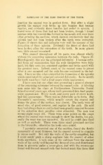
32 PATHOLOGY OF THE HARD TISSUES OF THE TEETH.
junction the enamel was in perfect form. But after a slight
growth the enamel rods broke up into bundles that became
smaller, and evidently these bundles had ended in spiculae. I
found none of these that had not been broken, though I found
patches with the mucoid film formed in the mouth still over them
after grinding the sections, which showed conclusively that the
spiculae had not been broken after the teeth were extracted.
Figure 38 shows this and indicates very well the manner of the
formation of these spiculae. Evidently the finest of these had
been broken after the extraction of the teeth. In many places
very little enamel remained.
This enamel throughout all its parts was almost wholly
without the cementing substance between the rods. Figure 39.
Histologically, this was the principal deformity. I became satis-
fied from my examination that the rods themselves were fully
hard, but they were not cemented together and broke apart with
the greatest ease. Indeed, much of the enamel came to pieces
after it was mounted and the rods became scattered in the bal-
sam. I have no idea what controlled the formation of the spiculae
which constituted the principal outward deformity. In the mouth
the teeth must have had a dead paper-white appearance.
I have seen but one other case presenting a general absence
of the cementing substance between the enamel rods. A laboring
man came into the clinic at Northwestern University Dental
School several years ago, whose teeth presented this dead paper-
white appearance. Every tooth, and every part of every tooth,
had this appearance. There was no deformity as to form. But
Naysmith's membrane, which usually covers the enamel and
forms the glaze of the surface, was absent. The teeth were of
usual size, of good contour, and regular in the arch. He said
they had always been so and he had been greatly annoyed because
of the attention their peculiar color attracted. The man was
twenty-eight years old. There were some points on the cusps
where the enamel was worn enough to show the dentin, but gen-
erally the wear was not excessive. He said he could chew food
as well as anybody. There were three small proximal cavities
in the bicuspids ; otherwise the teeth were sound.
I partially excavated one of the cavities, found the dentin
apparently of usual firmness, but the enamel seemed to crumble
to pieces easily. Not only the walls of the cavity crumbled, but
I could easily push a sharp explorer into the enamel of other
teeth anywhere. I took some of the cuttings from the enamel
walls of the cavity well beyond the decayed area and distributed
them in glycerin under a cover-glass, and with the microscope
found well-formed enamel rods that looked much like those that