Page 226 - My FlipBook
P. 226
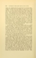
112 PATHOLOGY OP THE HARD TISSUES OP THE TEETH.
surface of a cuspid, which has progressed in the dentin, under-
mining the enamel, which, by breakage, has now formed a round
opening. At a time when this tooth was taking its place in the
arch and was uncovered by the gum tissue, only to about that
point, a decay penetrated the enamel and became seated in the
dentin. The particular form of the beginning in the enamel
is now lost by breakage from backward decay, but we know from
many observations of such cases that it was in a line across the
central portion of the present dark area. As the tooth pro-
truded farther through the gum, the conditions causing the
beginning of decay in the enamel passed away and did not return,
but the decay established in the dentin continued. Had the orig-
inal beginning failed, ever so little, to penetrate the enamel and
admit microorganisms to the dentin, the progress would have
ceased entirely. Later, a blackened blemish of the enamel would
have remained to show where decay had begun. Many of these
may be found in any box of extracted teeth.
A rare position of decay on the lingual surface of a cuspid
is shown in Figure 136, which, from what is left of the lingual
surface, seems to have begun as a pit decay, beginning, probably,
in pits at the junction of the lingual developmental grooves with
the linguo-gingival groove or pits along the lines of these. (See
"Descriptive Anatomy of the Human Teeth"— Black, Figures
5 and 20.) These grooves are unusually prominent, and one
undecayed pit remains in view. The case shows how decays may
sometimes begin in the most unusual positions and how difficult
it may become to define the local conditions causing them after
the immediate surroundings have been destroyed.
The gingival third decays in the buccal surfaces of the
bicuspids and molars are not materially different from labial
surface decays. They exhibit similar characters in both their
beginning and in their disposition to spread along the gum
margin mesially and distally. In the photograph of the upper
molar, Figure 137, two considerable areas of loss of enamel rods
appear in an area of rather faint whitening stretching across
the buccal surface. Figures 138, 139, photographs of a third
molar, show the wild race of destruction that sometimes befalls
these teeth when caries is allowed to go on unchecked by any
sort of cleaning. For these illustrations, the cementum has
been tinged with a selective anilin stain to show the gingival
line distinctly in order to bring prominently into view that por-
tion of the enamel covered by the free border of the gum, illus-
trating its protection from the beginnings of caries. This