Page 229 - My FlipBook
P. 229
CARIES AS A WHOLE. ITS CLINICAL FEATURES. 113
appears to Dest advantage in Figure 138. In Figure 139 it is
seen that the decays beginning in the mesial and the buccal sur-
faces have become connected across the mesio-buccal angle of
the tooth by a comparatively narrow neck. The pictures illus-
trate the riotous progress sometimes seen in buccal decays
under exceptionally unfavorable conditions. Figure 140 exhibits
another neglected buccal decay, which is a more ordinary exam-
ple of the form and extent of these when they are left to take
their own course. It will be noticed that this has not passed the
angles of the tooth. These decays will, of course, burrow along
the dento-enamel junction, the same as others, and in that way
destroy the enamel by backward decay to the gingival line, allow-
ing the free margin of the gum to fall into the cavity.
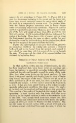
Figure 141, a photograph of an upper third molar, presents
an anomalous condition. In coming into position it deviated
backward and to the buccal from the normal, and seemed to
have stood for some time with but a part of its crown through
the gum. When removed, the whole of its exposed surface was
whitened by beginning decay.
Spreading of Decay Around the Teeth.
ILLUSTRATIONS: FIGURES 142-145.
In what has been written thus far of dental caries, the idea
has been developed that, when decay occurs on proximal sur-
faces, the tendency to superficial spreading is from the starting
point both buccally and lingually toward the angles of the teeth.
Also, that, when caries begins on the buccal surfaces, the ten-
dency is to spread mesially and distally from the place of begin-
ning toward the angles of the teeth. This is true of caries in
these positions in all of the teeth, but more especially of the
bicuspids and molars. A fewer number occur in the front teeth
as well. It has also been stated that in a few instances under
specially unfavorable conditions this decay crosses the angles
of the teeth and the proximal and buccal decays join each other.
This crossing of the angles of the teeth is the rarest of all of the
spreading. It then requires only that decay shall also occur
similarly on the lingual surfaces in order to complete the circle
of the tooth. This, though much more rare, occurs also. This
appears in the case of the lower second molar tooth, four photo-
graphs of which are shown in Figures 142-145, inclusive. Fig-
ure 142 shows the mesial surface with a broad, whitened area
of carious enamel stretching from angle to angle, in which the