Page 223 - My FlipBook
P. 223
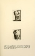
Figs. 138, 139. Two photographs of a lower molar tooth, in which caries proceeding- from a
buccal surface decay and a mesial surface decay have met across the angle. Figure 138 shows the
wide range of the decay of the buccal surface. Figure 139 shows the mesial surface decay as a broad,
whitened area, from which no enamel rods have fallen away. This meets the buccal surface decay
across the angle of the tooth. This picture also shows the breaking away of the undermined enamel
of the buccal surface to advantage. The cementum of this tooth was stained selectively with an anilin
dye to bring the gingival line into prominence, showing the influence of the free border of the gum in
protecting the enamel from beginnings of caries.