Page 218 - My FlipBook
P. 218
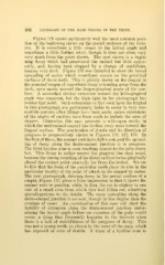
108 PATHOLOGY OF THE HAED TISSUES OF THE TEETH.
Figure 129 shows particularly well the most common posi-
tion of the beginning caries on the mesial surfaces of the incis-
ors. It is sometimes a little closer to the incisal angle and
sometimes a little farther away, though it does not often vary
very much from the point shown. The spot shown is a begin-
ning decay which had penetrated the enamel but little appar-
ently, and, having been stopped by a change of conditions,
became very dark. Figure 130 was intended to show the broad
spreading of caries which sometimes occurs on the proximal
surfaces of these teeth. This is plainly shown on the lingual in
the rounded tongue of superficial decay extending away from the
dark, open cavity toward the linguo-gingival angle of the sur-
face. A somewhat similar extension toward the labio-gingival
angle was apparent, but the high light in the photograph has
hidden that point. Such extensions as that seen upon the lingual
in this photograph are particularly liable to occur in very sus-
ceptible persons after fillings have been made, unless extensions
of the angles of cavities have been made to include the area of
danger. Otherwise, this case presents a wide-open cavity in
which the undermined enamel has broken away most toward the
lingual surface. The penetration of dentin and its direction of
progress is progressively shown in Figures 131, 132, 133. In
the first of these, the enamel rods have fallen out, and the spread-
ing of decay along the dento-enamel junction is in progress.
The faint hyaline zone is seen reaching almost to the pulp cham-
ber. This decay is rather nearer the gingival line than usual,
because the strong rounding of the distal surface inciso-gingivally
placed the contact point unusually far from the incisal. We see
in this that the form of the particular tooth plays its role in the
particular locality of the point of attack in the enamel by caries.
The next photograph, showing decay in the mesial surface of a
cuspid, Figure 132, gives a false impression in that it shows the
enamel rods in position, while, in fact, the cut is slightly to one
side of a small area from which they had fallen out, admitting
microorganisms to the dentin. The same spreading along the
dento-enamel junction is present, though in less degree than the
average of cases. An examination of this case will show the
liability of extension along the dento-enamel junction under-
mining the incisal angle before an exposure of the pulp would
occur, a thing that frequently happens to the incisors when
there is a lack of watchfulness of the progress of decay. This
was not a young tooth, as shown by the wear of the cusp, which
has exposed an area of dentin. A trace of a hyaline zone is