Page 374 - My FlipBook
P. 374
Fla. 203.
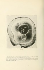
Flos. 20.1, 204, 205. A series of photomicrogr.aplis of cross sections of ;i iiuilar tooth showing
a carious cavity, prepared cavity, and a filling, in the distal surface portion.
Fio. 203. This shows the various decays. The decay in the distal surface has passed through
the enamel and entered the dentin ; there is a broad beginning of decay in the enamel of the mesial
surface. The section has cut across the deeper portion of a decay that has entered the denlin from
the central pit of the occlusal surface. The irregular white spot in the center is the result of an
injury to the photographic film.