Page 371 - My FlipBook
P. 371
EXCAVATION OF CAVITIES BY CLASSES. 165
of most of the portion of tlio I)UO('al dentin wall that is i)arailel
with the lingual dentin wall, but this could well be done by cut-
ting a little deeper convenience point to hold the filling during
the building of this portion. Notice particularly that if this were
a filling for a person fourteen years old, the recessional lines of
the pulp would have to be avoided for safety against pulp expos-
ure when forming the step. The dentin walls would have to be
converged to meet the narrower step.
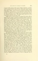
Figure 203 is a cross section of an upper first molar with an
injury by decay which is just penetrating the enamel in the distal
surface and extending much to the buccal because of a little irreg-
ularity of its position in relation to the second molar. The
greater part of the carious enamel has been lost. In this case
there was also an extensive pit decay in the occlusal portion, of
which the deeper part of the softened dentin is cut through and
has cracked in drying. This serves to obscure the position of the
recessional lines of the horns of the pulp. Notice also that a
decay which is broad from buccal to lingual was starting in the
enamel of the mesial surface and try to make out how broad
the cavitj^ in that surface would need to be to render it safe
against further decay. Immediately this question is asked, one
wishes to see the form of the contact with the next tooth, without
which the question can not be definitely answered.
The decayed enamel of the distal surface, the rods of which
had not fallen out in grinding, were accidentally lost in mount-
ing. The remains of enamel in such decays are often very frail.
There is practically no injury to the dentin. If all cavities of
decay could be filled thus early, it would be much better for the
teeth. The injury by decay of dentin to the depth of such a cav-
ity as shown in Figure 197 is a much greater injury to the tooth
than the cutting of such a cavity in sound dentin. In the latter
case, examinations have shown that the dentinal fibrils generally
remain alive between the filling and the dental pulp, while, if
the dentin has been decayed, they generally will be found dead
—
to the pulp chamber some years later. The form of cavity
omitting the convenience points — recommended for this case
is shown in Figure 204. In all cavities of the bucco-lingual
breadth shown in these series of cross sections, it is best to cut
the enamel walls and the dentin walls on distinctly different
planes as shown. In many of the wider extensions, this becomes
impracticable ; and then all or a part of the dentin wall parallel-
ing the opposite wall must be sacrificed in order to make the
extension sufficient for the purpose, and also bring the step as
23a