Page 379 - My FlipBook
P. 379
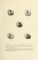
Fig. 206. Kio. 207.
Fig. 20
Fig. 209. Fio. 210.
Figs. 206-210. A series of pictures for the further illustration of cavity preparation in the
first molars.
Fig. 206. An upper first molar with a cavity in the mesial surface that has burrowed in the
dentin, but not sufficiently to require a very broad cavity for the complete removal of decay.
Fig. 207. A cavity prepared with the cutting as little extended as complete removal of all
undermined enamel would allow.
Fig. 208. The filling placed in the narrow cavity.
Fig. 209. The same tooth with a cavity prepared with the full limit that e.\tension for pre-
vention could allow in any case. Only actual undermining of enamel and destruction of dentin would
justify further extension. Generally so broad an extension could not be justified. A much less
extension than shown is very generally sufRcieiit. The narrow cavity, however, is too narrow to serve
in the usual conditions in such a case.
Fig. 210. The filling in the extended cavity.