Page 257 - My FlipBook
P. 257
Fig. IIG.
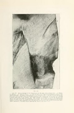
Fig. 116. Photomicrograph of the decayed area on the right side in Figure 114. d. Dentin.
E. Enamel. x. Area of decay. z. An extension of the superficial beginning of decay of the
enamel occlusally. The dento-enaniel junction is seen between d and e. In this case the enamel rods
have fallen into a tangled mass in the deeper portion of the central part of the decay at X, leaving
a partly open cavity in the enamel. Tlie dentin beneath is considerably decayed but has not been
pulled away from the enamel by shrinkage. The extension of the decay, as seen in the perpendicular
section, has been toward the occlusal portion of the tooth. The extension of beginning on the surface
seen at z is in a degree separated from the principal area of decay and extends toward the dento-
enamel junction in a flamelike tongue, following the course of the enamel rods.