Page 255 - My FlipBook
P. 255
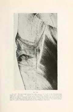
Fig. 115.
Fig. 115. Pliotomicrograph showing the carious area seen on the left of the small photograph,
Figure 114. d. Dentin. e. Enamel. x. Area of decay. y. Line of actual solution of the
calcium salts of the dentin. '/,. Uackward decay of the enamel, which shows very white by reflected
light, but is dark by transmitted light. In the drying of the specimen, the decayed dentin has
shrunken and pulled a little away from the enamel. A slight break of the t-naTiiel rods has occurred
at X, and a little confusion of the decayed rods has occurred near Ihe letter y. No enamel rods have
fallen out, however, and niicrotirgnnisms have not been admitted to the dentin.