Page 247 - My FlipBook
P. 247
Mm i
FlQ. 111.
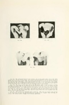
Fig. 109. This photograph discloses tliree decays; one in the mesial surface, one in the distal
surface, ami one in the occlusal surface. Tlie decay in the distal surface is not well sliown because of
the loss of a part of the enamel. The decay in the mesial surface is cut to one side of its central
area and shows the undermining of the enamel buccally beyond the area of penetration of the enamel,
and shows well the area of whitened bacliward decay of the enamel. The extraordinary prolongation
of the mesiobuccal horn of the pulp is also an interesting feature of the specimen.
Fig. 110. The decay on the right side of the illustration shows a very prominent clouding
extending to the pulp. This also shows particularly well the form of the clouded area, with the base of
the cone placed diagonal to its length, caused by the relation of the direction of the dentinal tubules
to the proximal surfaces. The actual decay of the dentin in this case is marked by the very dark
area about the small opening in the enamel. The spreading along the dento-enamel junction is very
wide. The decay in the mesial surface has exposed the pulp before the mesial marginal ridge has
broken so as to expose the cavity.
FiQ. 111. This tooth has an unusually heavy enamel cap. This seems not to have been any bar
to the penetration of caries, but has prevented the breaking of the marginal ridge disclosing the
cavity to the occlusal surface, though it is extensively undermined.