Page 245 - My FlipBook
P. 245
Fig. 108.
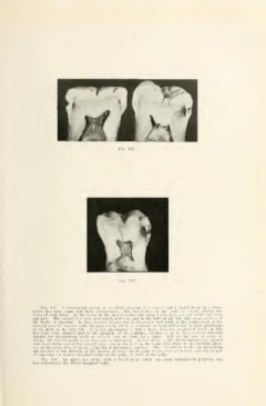
Fig. 107. A pliotograph giving an excellent showing of a mesial and a distal decay in a lower
molar that have made but little advancement. The two halves of the tooth are shown, giving two
views of each decay. In the decay in the mesial surface the enamel rods have not yet fallen out from
any part. The enamel has been penetrated, however, and in the half on the left side some solution of
the dentin is apparent. In this, another feature not so frequently met with, is the compression of the
decayed area by contact with the next tooth, which is apparent in both halves but is most prominent
is not uncommon
in the half on the left side. It to find a decay that has progressed about as this
has done (but situated just to the gingival of the contact), swollen so as to have become flattened
against the proximating tooth or even to take its form for a space. But for the area of decay to
occupy the contact point as in this case is infrequent. In the decay in the distal surface, the enamel
rods have fallen out of the central area, and in the half on the right side, there is an excellent show-
ing of the projection of decay along the deiito-enamel junction. The illustration is also an interesting
one because of the showing of the unusual nearness of the pulp Ui the occlusal surface and the danger
of exposing the mesial marginal ridge of the pulp, or horn of the pulp.
Fig. 108. An upper first molar with a distal decay which has made considerable progress, ana
has undermined the distal marginal ridge.