Page 58 - My FlipBook
P. 58
56 EMBRYOLOGY OF THE DENTAL TISSUES.
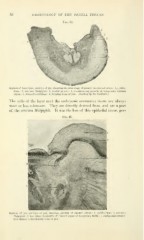
Fig. 34.
.Hf^"
Section of lower jaw, embryo of pig, sliowing lliu lirsl i^tage of growth in enaniel organ : 1, epithe-
lium 2, stratum Malpighii ; 3, dental groove ; 4, commencing growth of temporary enamel
;
organ ; 5, Meckel's cartilage ; 6, forming bone of jaw. (Section by Dr. Sudduth.)
The cells of the layer next the embryonic connective tissue are always
more or less columnar. They are directly derived from, and are a part
of, the stratum Malpighii. It was the loss of this epithelial tissue, per-
FiG. 35.
Section of jaw, embryo of pig, showing growth of enamel organ: 1, epithelium: 2, stratum
Malpighii : 3, first stage in growth of enamel organ of temporary tooth ; 4, embryonic connec-
tive tissue ; 5, developing bone of jaw.