Page 154 - My FlipBook
P. 154
;
152 DENTAL HISTOLOGY AND OPERATIVE DENTISTHY
membrane, they become less nuiiicroiis as age advances. These struct-
nres are specially well shown in the membranes of the pig and sheep.
Fig. 134 siiows their appearance in a transverse section of" the root of an
incisor of a sheej) ; here they swing out from the surface of the (lemen-
tnm and l)aci< again in looj)s, winding in and out among the fibers.
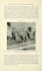
Studied witii higher powers (Fig. 1'35), they are seen t<» be made up of
epithelial cells, with large oval nuclei and reacting to tiie characteristic
epithelial stains. Tliey are arranged in cords, thftugh sometimes what
seems to be a lumen of a gland tubule can be found (Fig. 136). The
Fi(i. 136.
^a yiirrwr^fc'*.
Epithelial structures: Ec, epitliclial cord, M|iiiarcntly showing a lumen; b, cementoblasts
Cm, cementum ; D, dentin. (About 500 X.)
cords are invested with a delicate basement membrane, but no special
relation to bloodvessels has been demonstrated. The attempt to show
their connection with the surface epithelium has thus far failed. As the
gingivus is approached (Fig. 137), they seem to swing out from the sur-
face of the root and are lost between the projections of the epithelium
lining the gingival space. There is evidence that these structures are,
at least in some cases, of importance as the primary seat of pathological
conditions of the membrane.
152 DENTAL HISTOLOGY AND OPERATIVE DENTISTHY
membrane, they become less nuiiicroiis as age advances. These struct-
nres are specially well shown in the membranes of the pig and sheep.
Fig. 134 siiows their appearance in a transverse section of" the root of an
incisor of a sheej) ; here they swing out from the surface of the (lemen-
tnm and l)aci< again in looj)s, winding in and out among the fibers.
Studied witii higher powers (Fig. 1'35), they are seen t<» be made up of
epithelial cells, with large oval nuclei and reacting to tiie characteristic
epithelial stains. Tliey are arranged in cords, thftugh sometimes what
seems to be a lumen of a gland tubule can be found (Fig. 136). The
Fi(i. 136.
^a yiirrwr^fc'*.
Epithelial structures: Ec, epitliclial cord, M|iiiarcntly showing a lumen; b, cementoblasts
Cm, cementum ; D, dentin. (About 500 X.)
cords are invested with a delicate basement membrane, but no special
relation to bloodvessels has been demonstrated. The attempt to show
their connection with the surface epithelium has thus far failed. As the
gingivus is approached (Fig. 137), they seem to swing out from the sur-
face of the root and are lost between the projections of the epithelium
lining the gingival space. There is evidence that these structures are,
at least in some cases, of importance as the primary seat of pathological
conditions of the membrane.