Page 153 - My FlipBook
P. 153
PERIDENTAL MEMBRANE. 151
Black, ill his work on the periosteum and peridental membrane, in 1887,
and were called by him the glands of the peridental membrane. About
the same time von Brunu ' described what are probably the same struct-
nres, and which he regarded as embryonal remains of the inner layer
of the enamel organ, which he described as growing down over the sur-
face of the root. These structures appear as cords of epithelial cells
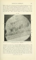
Fig. 135.
Epitlu'lial structures ^from sheep): /V*, tihrdMasts ; AV, epitliolial structures; r^, cenientiiblasts;
C'/n, cementum ; />, deutiu. (About 700 X.)
arranged in the form of a network winding between the fibers of the
membrane, very close to the cementum and surrounding the root almost to
the apex. Their arrangement is illustrated in Fig. 133, a diagram by Dr.
l^lack. The meshes of the net are close in the gingival portion of the
membrane, but grow more and more open in the alveolar portion. They
are not confined to the membranes of young teeth or the temporary den-
tition, as Dr. Black has shown them in the membrane of a tooth from
a man seventy years old, though, like all of the cellular elements of the
' Archivf. mikros. Anal , 1887.