Page 145 - My FlipBook
P. 145
PERIDESTAL MEMBRANE. 143
erupted tooth, and comparatively small and few in the membrane around
an old tooth. This is characteristic of fibroblasts in other positions.
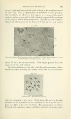
The fibroblasts are shown as they appear in a hematoxylin-stained
section with low powers in Fig. 124, which gives part of the membrane
in the gingival portion between two teeth. The cells are seen as spindle-
shaped dots which mark out the fibers ; at F they are seen in a position
Fig. 124.
Fibers and fibroblasts from transverse section of membrane : F, fibers cut transversely ; P", fibers
cut longitudinally, showing fibroblasts. (About 80 X.)
where the fibers are cut transversely. AVitli higher powers these cells
appear as in Figs. 126 and 135.
The cementoblasts are the cells which form the cementum, and are
found everywhere covering the surface of the root between the fibers
Fig. 125.
Cementoblasts. (Drawing by I)r. Black.)
which are imbedded in the tissue. While these cells perform the same
function for the cementum as the osteoblasts do for bone, they are in
form very different from the osteoblasts. The cementoblasts are always
flattened cells, sometimes almost scale-like, and when seen from above