Page 140 - My FlipBook
P. 140
138 DENTAL HISTOLOGY AND OPERATIVE DENTISTRY.
thoy form tlio ])rincipal bulk of the tissue, but they also perform the
principal function of the membrane, the support of the tooth and sur-
rounding tissues. The interfibrous tissue, also of the white variety but
made up of smaller and more delicate fibers, is found filling spaces
between the principal fibers and surrounding and accompanying the
bloodvessels and nerves.
For convenience of description and study, the peridental membrane
is divided into three portions : the(/inf/iva/, that portion which surrounds
the root occlusally from the border of the alveolar process ; the alveolar^
the portion from the border of the })rocess to the apex of the root ; and
Fi(i. ]'20.
D.
N.
F.
C.
^ w^;
/,
/.'.
M
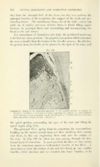
Longitudinal section
to the outer layer of the periosteum over the alveolar process ; F^, fibers attached to the bone
at the rim of the alveolus ; B, bone. (About 30 X-)
the apical portion, surrounding the apex of the root and filling the
apical region (Fig. 118).
The principal fibers spring from the cementum, the cementoblasts
building up the matrix around them and then calcifying both matrix
and fibers, in this way implanting their ends into the surface of
the root. In Fig. 119 the fibers are seen passing through the last-
formed layer of cementum. In most positions the fibers as they spring
from the cementum appear as well-marked bundles of fine fibers. A
short distance from the surface of the root they break up into smaller
bundles, Mdiich interlace and are reunited into larger buwdles, to be