Page 131 - My FlipBook
P. 131
PULP. 129.
layer of Weil the connective-tissue cells are especially numerous and
form a more or less distinct layer of closely placed cells. In the rest
of the body of the pulp the cells are about uniformly distributed through-
out the intercellular substance. These connective-tissue cells are of the
characteristic forms, rather small, containing a small l)ut deep-staining
nucleus, the protoplasm stretching out into slender projections in two
directions to form the spindle cells, or in more than two directions to
form the stellate cells. The stellate forms are more common in the
body of the pulp, the spindle form in the canal portions. The round
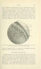
Fig. 111.
"^W
Odontoblasts and forming dentin: E, forming enamel; I), forming dentin; 0, odontoblasts;
Dp, body of dental papilla. (From photomicrograph by Rose.)
cells are comparatively few in number, and are probably young cells
which have not yet acquired the adult form.
BLOODVESSELS OF THE PULP.
The blood-supply of the pulp is extremely rich, several arterial ves-
sels entering in the region of the apex of the root, often through several
foramina. These large vessels extend occlusally through the central portion
of the tissue, giving off many branches which break up into a very close
and fine capillary plexus (Fig. 112). From the capillaries the blood is
collected into the veins, which pass apically through the central portion
of the tissue. A very striking peculiarity of the bloodvessels of the pulp
is the thinness of their walls. Even the large arteries show scarcely any