Page 130 - My FlipBook
P. 130
128 DENTAL HISTOLOGY AND OPERATIVE DENTISTRY.
the surface; but
in this section tlie cut was not in the (Urection of the
h)no: axis of tlie odontobhists, hut obliquely tliroutih
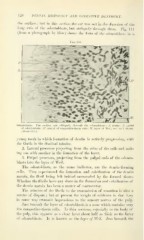
tiicin. Fij^. Ill
(from a photograph by Rose) shows the form of the odontoblasts
in a
Kk;. 110.
Odontoblasts. The section cuts obliquely through the odontoblasts : F, fibrils : .V, nuclei
of odontoblasts; N', nuclei of connective-tissue cells; W, layer of Weil, not well shown.
(About 80 X.)
young tooth i?i which formation of dentin is actively progressing, with
the fibrils in the dentinal tubules.
2. Lateral processes projecting from the sides of the cells and unit-
ing one with another in the formation of the layer,
3. Pulpal processes, projecting from the pulpal ends of the odonto-
blasts into the layer of Weil.
The odontoblasts, as the name indicates, are the dentin-forming
cells. They superintend the formation and calcification of the dentin
matrix, the fibril being left behind surrounded by the formed tissue.
Whether the fibrils have any share in the formation and calcification of
the dentin matrix has been a matter of controversy.
The relation of the fibrils to the transmission of sensation is also a
matter of dispute ; but at present the weight of evidence is that they
in some way transmit impressions to the sensory nerves of the pulp.
Just beneath the layer of odontoblasts is a zone which contains very
few connective-tissue cells. In thin sections, especially in the body of
the pulp, this appears as a clear layer about half as thick as the layer
of odontoblasts. It is known as the layer of Weil. Just beneath the