Page 95 - My FlipBook
P. 95
BONES. 105
The Anterior Border.—(For description see Coronoid Process.)
The Posterior Border at its upper portion is smooth and rounding.
As it approaches the angle of the bone it is roughened for the insertion
of the stylo-maxillary ligament.
Development.—The inferior maxilla is the second bone developed,
the clavicle being the first ; it is developed from the first pair of what
are known as the visceral or branchial folds or arches of the embryo,
called the uTandibular plates. These plates from the twenty-fifth to the
twenty-eighth day of embryonal life advance from the sides of the base
of the cranium and meet in the median line. Soon after this union
the cartilage of Meckel appears in the deeper portion of the mandibular
plate. In mammals the proximal end of this cartilage forms the mal-
leus (one of the small bones of the middle ear), and its distal portion
advances along the mandibular plate until it meets its fellow of the
opposite side at the symphysis menti.
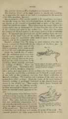
INIeckel's cartilage (Fig, 50) forms in great measure what may be
termed a temporary framework for the support of the lower jaw. It
disappears at the latter part of the
fifth or beginning of the sixth month Fig. 50.
of foetal life, and ossification proceeds.
About the fortieth day of embry-
onic life ossification commences from
several centres deposited on the out-
side, about midway between the prox-
imal and the distal extremities, in the
membrane which partially surround
Internal Face of the Right Maxilla of a
the cartila";e of Meckel. These cen Human Embryo of about Three Months,
showing the naturnl size atid the relative
tres speedily unite. Ossification then position of Meckel's cartilage.
proceeds in both directions along the
outer, under, and inner surface of the cartilage, but doss not unite with
About the sixtieth day a miniature jaw is formed, a siuall portion
it.
of the body at the symphysis resulting from direct ossification of
Meckel's cartilage. The condyles and a portion of the rami are also
ossified from other cartilage. From the centre of the rami internally
Meckel's cartilage is prolonged backward to the glenoid fissure, and
thence to the middle ear. That portion which passes between the
temporal bone and the inferior maxilla
becomes surrounded by fibrous tissue and
forms the internal lateral ligament of the
jaw.
At birth osseous union between the lat-
eral halves of the bone has not taken place,
they being connected by fibro-cartilaginous
tissue. They unite, however, during the
first year, ossification commencing below
Thp Inferior Maxilla of a Foetus at
and extending upward, a trace only remain- about the Full Teriofl of Intra-uter-
ine Life. The two sides (o, /)) are
ing at the upper portion at the beginning separate.
of the second year. The body of the bone
is shell-like, open at the top, and contains the germs of the teeth. The
coronoid processes are large proportionately to the remainder of the