Page 90 - My FlipBook
P. 90
100 ANATOMY
Development.—The malar bone is developed in membrane from
two points of ossification, which appear abont the eighth week of
embryonal life, uniting about the fourth month. Occasionally the two
portions of the bone remain seimrate throughout life. When this is
the case the bone is divided into an upper and a lower portion by a
horizontal interspace, the upper portion being the larger.
The Inferior Maxillary Bone.
The importance of the inferior maxillary bone, mandible, or lower
jaw to the dentist and the surgeon cannot be over-estimated. Its posi-
tion, composition, and development, its nerve- and blood-supply, com-
bine to render it liable to various and grave diseases. In order that
these shall be thoroughly understood and properly treated, a detailed
knowledge of its anatomy is absolutely necessary.
The inferior maxilla is symmetrical in form, and is situated below
the alveolar border of the superior maxilla, beneath the zygomatic and
glenoid fossre, articulating in the latter cavity. The lower border,
extending from side to side, forms the anterior inferior boundary of
the face. It assists in forming the lateral portions of the outer bound-
aries of the zygomatic fossae. It also forms the greater portion of the
superior boundary of the surgical squares of the neck and the digastric
triangles.
The inferior maxilla is the largest, heaviest, and strongest bone of the
head, and contains one-half the teeth. It presents for examination a
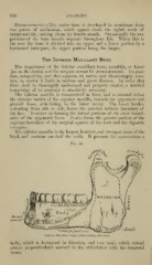
Fig. 48.
.V«-«/«Z ,,^^_ ..^_ _„
» & DEp-'-ANculi....?.?.'---' ^^f
/troc/xj ^4cp:Lflal|^•NV6.?"^::•;;.lUis'.•** S
uf
G foove J'o>- farinf arty
Inferior Maxillary Boue, outer surface, side view.
body, which is horizontal in direction, and two rami, which extend
almost perpendicularly upward to the articulation with the temporal
bones.