Page 681 - My FlipBook
P. 681
—
INFLAMiVA TION. 691
ihat are seen passing the field of view. Finally, some are seen to
become more decidedly adherent to the wall, as if fused with it, and
others to be likewise adherent in the neighborhood. As this progresses
they begin to be piled the one on the other ; and all this time the blood-
current is becoming slower and slower. Some of the white globules
that have seemed to hold fast are seen to loosen, and after swaying for
a time float away with the current. In all this movement it will be
noticed that the globules appear to be developing an adhesiveness that
they did not manifest at the beginning of the observation. Those that
gradually break away and move off from the focus of the irritation
Avhich now can only be seen in this disposition to stickiness—seem to
lose this property as they recede from the field. This will give the
impression that it is the vesseVs wall in which this stickiness is devel-
oped, and not in the blood-globule. In the focus of this action the adhe-
sion of the white o-lobules will o-q on until the entire inner wall of the
vessel is completely covered as with a pavement, and they may be piled
one upon the other. This adhesion of the globules is the first step in
the process that can be considered as significant of inflammation. It
may indeed be inferred that the hypereemia is that of irritation, and will
lead to inflammation ; but there is nothing in the microscopic appear-
ance of the tissues or of the blood in the vessels by which the diflerence
can be noted until the adhesion of the white globules has become man-
ifest. With the adhesion of the white globules, as it advances, there is
also seen a disposition to the adhesion of the red. These at first occu-
pied the centre of the blood-streams, but as the adhesion of the white
globules progresses the channels become narrowed, the motion of the
Fig. 384.
Fig. 385.
^0^ ^^
y.^'%r.^ J^h^
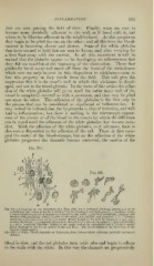
Fig. 3S4.—A Capillary of the iMesentery of a Frog nine hours inflamed, showing detachment of an
endothelial cell, which is finally carried off by the blood-current (high-power) : e, capillary walls;
/, white blood-corpuscles or leucocytes external to the walls; /, capillary endothelia, granular and
swollen with projecting liellies; ff, cells of adventitia, also swollen and granular; o, (/, /, colorless
corpuscles ailherent to the walls; d is rather firmly bound to the wall by means of a bud pene-
trating the latter ; /, a corpuscle adherent to the point of union of two adjacent endothelial cells;
a, a white corpuscle adhering tightly to the upper end of an endothelial cell, b, which is partly
pried out from its bed by the action of the red discs. The arrow indicates the direction of the
current (Shakespeare).
Fig. .385.— a White Blood-corpuscle, or Leucocyte, from human blood, showing amceboid movement
(Klein).
blood is slow, and the red globules turn aside also and begin to adhere
to the walls with the white. In this way the channels are progressively
INFLAMiVA TION. 691
ihat are seen passing the field of view. Finally, some are seen to
become more decidedly adherent to the wall, as if fused with it, and
others to be likewise adherent in the neighborhood. As this progresses
they begin to be piled the one on the other ; and all this time the blood-
current is becoming slower and slower. Some of the white globules
that have seemed to hold fast are seen to loosen, and after swaying for
a time float away with the current. In all this movement it will be
noticed that the globules appear to be developing an adhesiveness that
they did not manifest at the beginning of the observation. Those that
gradually break away and move off from the focus of the irritation
Avhich now can only be seen in this disposition to stickiness—seem to
lose this property as they recede from the field. This will give the
impression that it is the vesseVs wall in which this stickiness is devel-
oped, and not in the blood-globule. In the focus of this action the adhe-
sion of the white o-lobules will o-q on until the entire inner wall of the
vessel is completely covered as with a pavement, and they may be piled
one upon the other. This adhesion of the globules is the first step in
the process that can be considered as significant of inflammation. It
may indeed be inferred that the hypereemia is that of irritation, and will
lead to inflammation ; but there is nothing in the microscopic appear-
ance of the tissues or of the blood in the vessels by which the diflerence
can be noted until the adhesion of the white globules has become man-
ifest. With the adhesion of the white globules, as it advances, there is
also seen a disposition to the adhesion of the red. These at first occu-
pied the centre of the blood-streams, but as the adhesion of the white
globules progresses the channels become narrowed, the motion of the
Fig. 384.
Fig. 385.
^0^ ^^
y.^'%r.^ J^h^
Fig. 3S4.—A Capillary of the iMesentery of a Frog nine hours inflamed, showing detachment of an
endothelial cell, which is finally carried off by the blood-current (high-power) : e, capillary walls;
/, white blood-corpuscles or leucocytes external to the walls; /, capillary endothelia, granular and
swollen with projecting liellies; ff, cells of adventitia, also swollen and granular; o, (/, /, colorless
corpuscles ailherent to the walls; d is rather firmly bound to the wall by means of a bud pene-
trating the latter ; /, a corpuscle adherent to the point of union of two adjacent endothelial cells;
a, a white corpuscle adhering tightly to the upper end of an endothelial cell, b, which is partly
pried out from its bed by the action of the red discs. The arrow indicates the direction of the
current (Shakespeare).
Fig. .385.— a White Blood-corpuscle, or Leucocyte, from human blood, showing amceboid movement
(Klein).
blood is slow, and the red globules turn aside also and begin to adhere
to the walls with the white. In this way the channels are progressively