Page 546 - My FlipBook
P. 546
556 DENTAL EMBRYOLOGY AXD HISTOLOGY.
It surrounds the lower jaw, forming a lining for the mouth and an outer
coat for the jaw,
Tlie jaw is a process which has budded off from the main body of
the blastoderm, and is composed of a layer of mesodermic tissue sur-
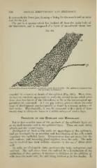
FiG. 303.
c(, coiiiiectivt; tissue ol" uiesoblast ; t-y;, ejiiblast single laj'pr of cells). The epiblast is separated from
the mesoblast mechanically.
rounded bv a yesicle or sheath of the e])iblast (Fig. 303). Here, then,
we have an excellent opportunity to study the several tissues A^'hich arise
from these two layers of the blastoderm, as far as the microscopical ap-
pearances are concerned. A 1 cm. pig embryo prc-^cnts about the same
stage of develojiment macroscopically as found in a human embryo of
four weeks. Histologically, it may be comjiared with a chick of from
twenty-four to thirty-six hours or a rabbit embryo of twelve days.
Products op the Epiblast and Mesoblast.
Let us first consider some of the products of the epiblastic layer, or,
as we shall hereafter call it, the epithelUd layer. These are nails, hairs,
glands, and the oiamcl organ.
Development of Kails.—The nails are appendages of the ejiidermis,
and are developed })y an accretion and hornification of the cells which
constitute the epithelial layer. Desquamation does not occur, but the
cells coalesce, and, liccoming glued together, form the nails. The nails
can be re.solved into their cellular elements by the use of dilute nitric
acid.
In nails we distinguish three portion,-;—the body, nail-groove, and
nail-bed. The nail arises from the nail-bed by a hornification of the
ejiithelium of that portion; it increases in thickness by the addition of
cells from the under side, the nail being thickest at its free border. It