Page 544 - My FlipBook
P. 544
554 DENTAL EMBRYOLOGY AND HISTOLOGY.
influence of the osteoblasts the cartilage is broken down and becomes
ossified and incorporated into the substance of the maxilla. These
changes are very plainly sho\vn in the accompanying cut, from a photo-
micrograpli (Fig. 300).
Ossification of Meckel's cartilage differs from that known as intercar-
til((gi)iou.s; in the latter case there is rapid proliferation of the cartilage-
cells, the cartilaginous head (femur) increasing in size in proportion to
the encroachment of the ossification zone. This does not occur in ossi-
fication of Meckel's cartilage. There is no increase of cartilage-cells,
except at the points of articulation, where true intercartilaginous ossi-
fication occurs. In the body of the jaw tlie cartilage simply becomes
calcified, and afterward ossifi(Kl and incorporated into the substance of
the maxilla, as before stated. It entirely disappears before the fifth
month—not by icasfing away, but by ossification. This change begins,
as we have seen, at two and a half months ; at three months it is almost
complete, and at four months, in nearly every case which I have exam-
ined, no trace of the cartilage remains. In the pig it persists much
longer, and is unaffected by ossific processes in embryos ten centimeters
in length.
Development of the Blastoderm.
In the unincubated chicken egg the blastoderm is composed of two
layers—the epiblast and the hypoblast. There is little or no change
observable until after the eighth hour of incubation, but between this
and the twelfth hour marked changes occur. Cross-sections of the
chick at this time will show a decided proliferation of the epiblastic
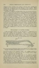
Fig 301.
-ep
Transverse Section through a Blastoderm of Chick, about the eightli hour after incubation (after Bal-
four); the section passes througli the middle of the primitive streak: juis, primitive streak;
ep, epiblast: hy, hypoblast; yk, yolk of the germinal wall.
layer in the region of the primitive streak. The epiblast, previous to
this time, is fi)rmed of a single layer of cells somewhat oval in shape,
i\\\i\ to the lateral pressure of fellow-cells.
The hy]iol)last consists of a single layer of cells, which lie parallel
witii the surface of the blastoderm, while the epiblastic layer stands,
palisade-like, upon the surface. Between the flat layer of the hypoblast
and the proliferated cells of the epiblast, are seen a few scattering cells
which appear to have arisen from the hypoblast, but the greater part
of the thickening is from the epiblast.
There is little or no change appreciable in the histological appearance
of the blastoderm until about the eighteenth or twentieth hour, when the