Page 538 - My FlipBook
P. 538
548 DENTAL EMBRYOLOGY AND HISTOLOGY.
ently appear. Previous to the formation of the latter, however, there
is formed a third layer, which locates itself between those already
developed, and is known as the mesoblast. It is mainly produced by
the proliferation of the cells of the epiblast (Fig. 292).
The cellular activity of the epiblast proceeds rapidly, and results in
the formation of two medullary plates which arise in parallel rows,
between which lie the medullary groove or primitive streak. At the
anterior portion, in the very first differentiation of the groove, a dark
spot is seen, known as the '' nodal point of Hensen," which subse-
quently marks the front part of the groove. Its signification is not
exactly known.
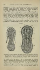
The medullary plates develop rapidly, expanding at their anterior
portion into a spatula-shaped crescent (Fig. 293). This gives rise to
Fig. 293.
^mmmm^^"^
\iiil)|"M" iiiiii.""iiiilwiii'
h
'f
^.,_
Rabbit Embryos of about tbe Ninth Day, seen from the doisal side (from Kcilliker) : «6, optic vesicle;
nf, amnion ; "/), area pellueida; A and hz, heart: hf, lueduilary plate in region of future fore-brain
;
/('", medullary plate in region of future mid-brain
; ///( and Ii"\ hind-brain ; vth. mid-brain ; ]ili,
pericardial section of body-cavity //,-, lateral zone; pr, primitive streak; ;/, medullary groove;
;
.sir, vertebral zone; iiu\ protovertebne ; vh, fure-brain , ro, vitelline vein.
the ce]ihalic end of the embryo. The first indication of the vertebral
column is seen about the eighth day, in the formation of the first pair of
somites. Tlicy are located in the region of the neck, and mark the line
of union of head and trunk. The latter gradually elongates by the
addition of other pairs of somites, the growth in length being from the
first-formed somites caudal-ward. The medullary groove deepens as