Page 534 - My FlipBook
P. 534
DENTAL EMBRYOLOGY AND HISTOLOGY
544
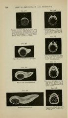
Fig. 284.
Fig. 280.
Diagram of Frog's Egg, in an Early .Stage Cross-section of Frog's Egg,
showing blastoderm same age
of IJevelopuient, longitudinal section: 1, a, a, lateral folds
thickened portion of external blas^todermic as I-Mg. 280:
situated upon^ either side of
layer; 2, anterior extremity of the embryo;
3, "posterior extremity, 4, internal blasto- groove B.
dermic layer; 5, cavity of vitellus.
Fig. 285.
Fig. 281.
Cross-section of Frog's I'^gg, same
stage of development as seen
in Fig. 281 : a, a, lateral pro-
Egg of Frog iu Process of Development. cesses ; B, neural groove.
Fig. 286.
Fig. 282.
Cross-section of Tadpole, showing
Egg of Frog, farther advanced. same stage of development as
seen in Fig. 282: B, neural
canal ; v, c, lateral processes of
spinal column.
Fig. 287.
Fig. 283
Tadjiole, fully developed. Cross-section of Fully-developed
Tadpole : letters same as seen
in Fig. 286.