Page 440 - My FlipBook
P. 440
450 DENTAL ANATOMY.
with a principal hook-shaped cusp, a small posterior basal cusp, and a
strong internal cingulum.
The next three premolars are similar, except that they are larger,
implanted by two roots, and have two posterior accessory cusps, the
hindermost of which is very small. The single molar diliers from the
rest of the teeth in advance of it in having an anterior basal cusp, being
relatively thicker at the base of the crown, and with a moderately well-
defined internal cingulum, which displays a tendency to develop inter-
nal cusps.
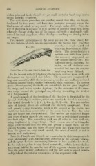
The incisors and canines of the lower jaw are like those above, but
the two incisors of each side are separated at the median line. The first
premolar is single-rooted and
somewdiat larger than its fellow
above. The crown displays a
median cone with three poste-
rior accessory cusps, and one
very minute anterior one. The
following teeth, including the
molar, are all similarly con-
structed, but have the anterior
Vertical View of the Lower Jaw of a Harbor Seal. , , i j.. ^ n a
basal cusp better denned.
In the hooded seals (Ci/stophora) the incisors are two upon each side
above, and one upon each side below. The canines are comparatively
large and powerful, while the molars and premolars are small and reduced
to simple conical bodies, similar to the teeth of the cetaceans. In another
genus (Stenorhynchii.s) the teeth are remarkable for the great length of
the cusps, and in one species, Lepfonyx, for the curvature of the acces-
sory cusps toward the principal one, thereby resembling the trident
of a fishing-spear.
A good example of the dentition of the Otaridce is furnished by the
fur seal {Callorhynns urs^nus),^\\\\Q\\ can usually be found in museums.
Tlie dental formula is I. f , C. }, Pm. f , M. = 36. The two median
f
pairs of incisors above are subequal and laterally compressed. They
each present a deep transverse notch in the summit of the crown, into
which the incisiform extremities of the lower incisors bite ; the outer
pair are larger and sharp-pointed. The canines are relatively longer
and more slender than in the seals, and have a well-defined posterior
trenchant edge. The succeeding teeth are all alike in form and size,
being imi)lanted by single fangs. Their crowns are of a triangular
shape when viewed from the side, and present a single cusp. It fre-
quently happens that the bases of these teeth just where they emerge
from the gums are very much eroded, the cause of which is not at
present well understood.
Both the PJiocidce and OtarirJcc are remarkable for their comparatively
weak and slender jaws, the backward dii-ection of the coronoid process,
and the great distance intervening between its base and the last tooth.
In the seals the palate is very broad posteriorly, and the last tooth does
not extend behind the anterior root of the zygoma, whereas in the sea-
lions the ]xalate is long and narrow, and the last tooth is placed consid-
erably behind the anterior termination of the zygomatic arch.