Page 28 - My FlipBook
P. 28
38 AXATOMY.
Fig. 3. vertebra will become like a sponge, and the long bones
may be tied into knots (see Fig. 3). In this ^\•ay the
internal parts of bone may be prepared for study by
cutting away portions with a sharp knife or pair of
scissors.
A preferable manner of preparing bone for microscopi-
cal examination is to take a small piece, about half a
cubic inch in size, of the compact portion of a long bone,
either of man, dog, cat, or rabbit, and immerse it in an
aqueous solution of chromic or picric acid, either of
which hardens the organic tissue as well as dissolves
the inorganic matter, and renders the tissue capable of
being cut into thin sections, Avhich are to be stained or
not according to methods in vogue by practical histol-
ogists. Hard sections can also be made by sawing a
small piece from a long bone and grinding it upon a
whetstone or plate of glass with emery-powder until
sufficiently thin.
When a bone is placed in a slow fire or a sufficiently
Fibula tied ^ heated furnace, the organic material will be consumed,
Knot after JIace
ration in j Bihife leaving oulv inorgauic substance and. the ash from the
men preserved in Organic matter. The shape is still preserved, but the speci-
spiiitA
j^gj^ jg ^,gj,^, brittle and will crumble almost at the touch.
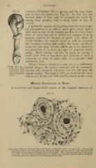
Minute Structure of Bone.
A transverse and longitudinal section of the compact structure of
Fig. 4.
Fransverse Section of rompact Tissue "f Humerus (mapnitied about InO diameters). Tbree of the
Haversian canals are seen, with their concentric rings; also the lacunie, with the canaliculi
extendinc from them across the direction of the lamellie. The Haversian apertures had become
tilled with ;iir and d'bris in grindinc down the section, and therefore appear black in the figure,
wh.ch represents the object as viewed with transmitted light.
Fig. 3. vertebra will become like a sponge, and the long bones
may be tied into knots (see Fig. 3). In this ^\•ay the
internal parts of bone may be prepared for study by
cutting away portions with a sharp knife or pair of
scissors.
A preferable manner of preparing bone for microscopi-
cal examination is to take a small piece, about half a
cubic inch in size, of the compact portion of a long bone,
either of man, dog, cat, or rabbit, and immerse it in an
aqueous solution of chromic or picric acid, either of
which hardens the organic tissue as well as dissolves
the inorganic matter, and renders the tissue capable of
being cut into thin sections, Avhich are to be stained or
not according to methods in vogue by practical histol-
ogists. Hard sections can also be made by sawing a
small piece from a long bone and grinding it upon a
whetstone or plate of glass with emery-powder until
sufficiently thin.
When a bone is placed in a slow fire or a sufficiently
Fibula tied ^ heated furnace, the organic material will be consumed,
Knot after JIace
ration in j Bihife leaving oulv inorgauic substance and. the ash from the
men preserved in Organic matter. The shape is still preserved, but the speci-
spiiitA
j^gj^ jg ^,gj,^, brittle and will crumble almost at the touch.
Minute Structure of Bone.
A transverse and longitudinal section of the compact structure of
Fig. 4.
Fransverse Section of rompact Tissue "f Humerus (mapnitied about InO diameters). Tbree of the
Haversian canals are seen, with their concentric rings; also the lacunie, with the canaliculi
extendinc from them across the direction of the lamellie. The Haversian apertures had become
tilled with ;iir and d'bris in grindinc down the section, and therefore appear black in the figure,
wh.ch represents the object as viewed with transmitted light.