Page 256 - My FlipBook
P. 256
266 ANATOMY.
the common sheath or epineurimii. Immediately beneath this sheath
are irregular lymph-spaces communicating with each other. A fibrous
layer, the perineurium, surrounds and forms a sheath for the different
bundles, giving room for the passage of blood-vessels supplying the
nerves. This layer is similar to the sheath of a muscle which forms a
covering to the bundles of muscular fibres. Within each bundle can
be seen the nerve-fibres, consisting of axis-cylinder, medullary sheath,
and neurilemma or sheath of Schwann, enveloped by a delicate tissue,
the endoneurium..
The Axis-cylinder (axial-band, axial-fibres) is the essential portion
it is nearly uniform in diameter, and undergoes no
of the nerve-fibre ;
interruption from the nerve-centre to near its peripheral distribution.
It is either cylindrical or flattened in shape, and passes nearly in the
central axis of the tube. AVhen in a fresh condition it appears pale and
transparent, and when examined with a microscope, using a high ])ower,
it is demonstrated to be composed of very fine homogeneous or more or
less beaded fibrilloe.
These elementary or primitive fibrilke of Max >SchuIfz are held together
by a faintly granular albuminous cement or interstitial substance. At
the termination of the axis-cylinder it is observed to divide up into
numerous fine filaments or fibrils. Some investigators claim that the
axis-cylinder has an independent or elastic sheath composed of neuro-
keratin.
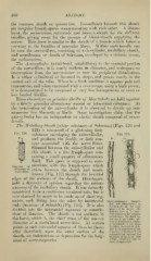
The Medullary Sheath (white substance of Schwann) (Figs. 120 and
121) is composed of a glistening fatty
Fig. 120.
substance enveloping the axis-cylinder, Fig. 121.
and produces the double or dark con-
tour associated with the nerve-fibres.
Situated between the axis-cylinder and
this sheath is a fine lymph-space con-
taining a small quantity of albuminous
fluid. This space is supposed to corn-
Diagram of municate with the lymph-space which
structure of exists betwceu the sheath and neuri-
lemma (Fig. 122) through the bevelled
edges of the sections of the sheath. Histologists
hold a diversity of opinion regarding the minute
anatomy of the medullary sheath. It was formerly
considered to be a continuous insulated tube, but is
now claimed by many to be made up of short seg-
ments, each fitting into the other by imbricated Kerve-substance (magni-
" "
" fied " 200 diameters) : a,
ends (incisions of Schmidt) (Fig. 123). It is also Kerve-tube of tlie com-
"
mon eel in water: tlie
divided into the internodal segments or constric-
delicate line on its exte-
tions of Ranvier. The sheath is not uniform in rior indicates the t ubular
membrane; the dark in-
thickness, which is the chief cause of the uneven ner one is the white sub-
stance of Schwann, si ijiht-
diameter of a medullated nerve-fibre. At certain ly wrinkled; b, the same
in ether. Several oil-glob-
points in each internodal segment of Ranvier (here-
ules have coalesced in the
after described), upon the outer surface of the interior, and others have
accumulated around the
sheath, are indentations or depressions for the lodg- exteriorof the tube. The
white substance has in
ment of nerve-corpuscles.
pai't disajjpeared.