Page 252 - My FlipBook
P. 252
262 ANATOMY
simi)ly tubes grooved in the boue, their lining membrane being com-
}30sed of pavement epithelium, with some elastic tissue between the
epithelium and the tubes. As they pass downward and join other tubes
they increase in size and their lining tissue becomes more and more
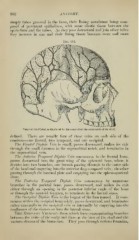
Veins of the Diploe, as displayed by the removal of the outer table of the skull.
defined. There are usually four of these veins on each side of the
cranium—one frontal, two temporal, and one occipital.
The Froiitd/ Diploic Vein is small, ])asses downward, makes its exit
through the small foramen in the supraorbital notch, and terminates in
the supraorbital vein.
The Anterior Temporal Diploic Vein commences in the frontal bone,
passes downward into the great wing of the sphenoid bone, where it
divides into two branches, one branch jiassing through to the outer side
of the head and emptying into the anterior deep temporal vein; the other
passing through the internal })late and emptying into the spheno-parietal
sinus.
The Poderior Temporal Diploic Vein commences by numerous
branches in the parietal bone, passes downward, and makes its exit
either through an o])ening in the posterior inferior angle of the bone
or through the mastoid fonunen, to terminate in the lateral sinus.
The ()ccij)i/(if J)iph)ic Vein is the largest of the four named. It com-
mences within the occipital boue solely, passes downward, and terminates
either externally in the occi]Mtal vein or internally by emptying into the
confluence of the sinuses or into the lateral sinus.
The Emissary Veins are those which form communicating branches
between the veins of the scalj) and those at tlie base of tlie skull and the
various sinuses of the brain-case. They pass through various foramina^