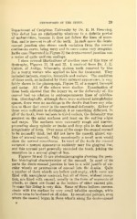
Page 79 - My FlipBook
P. 79
DYSTROPHIES OF THE TEETH. 29
Department of Creighton University by Dr. E. H. Bruening.
This defect has no relationship whatever to a definite period
of malnutrition, because it does not follow the lines of accre-
tion, and is present in all of the teeth. In such cases the dento-
enamel junction also shows much variation from the normal
continuous curve, being wavy and in some cases very irregular.
In the case illustrated in Figure 32 the dento-enamel junction was
a series of quite uniform scallops.
I show several illustrations of another case of this type of
dystrophy, Figures 33, 34 and 35. I received from Dr. J. E.
Callow, of Antigo, Wisconsin, sixteen teeth removed by him
for a young woman who applied to him for treatment. They
included incisors, cuspids, bicuspids and molars. The condition
of these teeth, as indicated by their outward appearance, is very
fairly shown in the photograph, Figure 33, of a cuspid, bicuspid
and molar. All of the others were similar. Examination of
these teeth showed that the injury to, or the deformity of, the
enamel had no relation to contemporaneous lines of calcifica-
tion. Histologically, although there were scattered interglobular
spaces, there were no markings in the dentin that bore any rela-
tion to those that occur in the accretional deformity. Either of
these were sufficient to distinguish it as something different. In
all of the teeth, from incisors to third molars, the deformity was
greatest on the axial surfaces and least on the cutting edges
and cusps. The surfaces were extremely rough and uneven,
presenting sharp spiculse or knobs and deep pits in the utmost
irregularity of form. Over some of the cusps the enamel seemed
to be normally thick, but did not have the smooth glazed sur-
face of normal enamel. Only occasionally a small area would
show the normal smoothness. In most of the teeth the enamel
assumed a normal appearance suddenly near the gingival line,
and this normal part generally encircled the tooth, joining the
cementum in a normal gingival line.
Figures 34 and 35 are photomicrographs showing the pecu-
liar histological characteristics of the enamel. In most of its
parts the dento-enamel junction is lost in a wild jumble of cir-
cular whorls or protrusions of enamel into the dentin. Quite
a number of these whorls are hollow and empty, while some are
filled with amorphous material, but all of these, without excep-
tion, are lined with enamel, usually in the form of segments of
whorls, as these are found in the bottom of other enamel pits.
In some this lining is very thin. Some of these hollows commu-
nicate with the surface by very small tubelike openings, while
others seem to be closed on all sides. In occasional patches, even
where the enamel began in these whorls along the dento-enamel