Page 76 - My FlipBook
P. 76
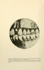
Fig. 32.— This photograph is from a skull found in the anatomical laboratory of the dental
department of Creighton University by Dr. E. H. Bruening. All of the teeth of this individual
presented the same deformity as those shown in the illustration.
A section prepared from this skull was lost by accident. The scalloping was very regular.
In this case the teeth presented an irregular wrinkling upon their surfaces, the wrinkles passing
horizontally around the teeth. These wrinkled teeth have always a scalloping of the dento-enamel
junction.