Page 50 - My FlipBook
P. 50
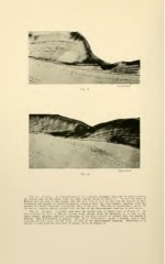
Fig. 13. Atrophy. A photomicrograph of a portion including the zone of injury nearest
the incisal edge on the labial from the same section shown in Figure 11. In this the lines of
Retzius may be seen in the enamel, also the dark line of junction between the enamel of first
formation and enamel of second formation, reaching from the dento-enamel junction, with the
enamel of second formation overlapping that of the first. The line of interglobular spaces in
the dentin, running almost parallel with the line of the dento-enamel junction, is well shown.
Fig. 14. Atrophy. A portion including the second zone of injury seen in Figure 11. In
this position the lines of Retzius diverge more sharply from the direction of the line of the
dento-enamel junction, and the overlapping of the third growth of enamel onto the second is
shorter. The discoloration is greater. The line of interglobular spaces is broader, and in this
position diverges more sharply from the line of the dento-enamel junction. Otherwise it is
similar in plan with the first zone of injury shown in Figure 13.