Page 45 - My FlipBook
P. 45
DYSTROPHIES OF THE TEETH. 15
now with the diagram shows that the growth has been arrested
on the lines of accretion or lines of Eetzius, as you may please
to call these lines, Figure 10, in both cases. Also, it is seen that
the second injury in Figure 11 is similar in plan to the first,
differing in detail only because of the changed direction of the
lines of accretion. In Figure 11 the incisal edge is broken, as
usually occurs in these thin edges, but Figure 12 is from a tooth
extracted soon after it came through the gums and all of the tis-
sue formed is present.
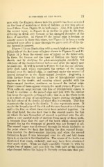
Figure 13 is an illustration with a much higher power of the
labial side of the first zone of injury shown in Figures 11 and 12.
Figure 14 is from the second zone of injury on the labial side.
In these, the tissues and the lines of Eetzius are fairly well
shown, and by studying the photomicrographs carefully, the
relations of the tissues formed before and after the injury may
be made out. It will be noted in Figure 13 that the one particu-
larly dark band, which represents the surface of the enamel
formed over the incisal edge, is continued under the enamel of
second formation to the dento-enamel junction. Beginning a
little farther from the incisal, a line of interglobular spaces
appears in the dentin, and running almost parallel with the
dento-enamel junction, continues on toward the incisal edge.
Faint traces of these appear even in the small picture, Figure 11.
"With sufficient amplification, this line of interglobular spaces is
found to continue to the incisal edge and join with the similar
line from the opposite or lingual side ; that is, in the whole tooth
it is a sheet or zone of interglobular spaces passing throughout
the full extent of the dentin, of which this is a section. This line
represents the injury in the dentin. It also represents more. It
marks the boundaries of the old and the new formation of dentin
and is the line on which these have been patched together. On
the other hand, the one dark line in the enamel marks the line
on which the new formation of enamel is patched onto the old.
After a very careful study of sections from many of these teeth,
it becomes clear that the part of the tooth which should have
formed during the stoppage of growth was not formed at all.
The enamel organ was destroyed through its whole thickness to
the point where the dark line limiting the first enamel forma-
tion reaches the dento-enamel junction, and when the second
formation began it was telescoped over the old and laid down
upon it, as shown in the illustration. The crown of the tooth was
shortened that much, certainly, and may have been shortened
very much more. When we study carefully Figure 12, with its
single line of injury, and note how the little part of the incisal
edge formed before the injury is literally sunken into that por-