Page 195 - My FlipBook
P. 195
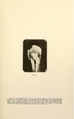
Fig. 114 The photograph in this case was taken with the surface dry, with the expectation that
the of decay would show whiter.
This succeeded well with the decay on the left side, but not
with the one right. The hyaline area of the left decay is well shown.
The extension occlusally
of decay along the dento-enamel junction and tin. very white backward decay of the enamel are inter-
,.
esting features.
After this photograph was made, the polished surface was cemented
cover glass
and ground thin for photomicrographing, and Figures 115, 116 were made.