Page 194 - My FlipBook
P. 194
98 PATHOLOGY OF THE HABD TISSUES OF THE TEETH.
great advantage. It will be noticed that in the cavity on the left
side of Figure 117, decay has already begun to undermine the
enamel forming the marginal ridge, and the distance to the pulp
is so great that the breakage of the marginal ridge would prob-
ably occur, disclosing the presence of the cavity before the pulp
would become involved. But this tooth shows that the pulp has
receded and is smaller than usual. In many cases the pulp is
involved before the breakage of the marginal ridge. This brings
us to the necessity of discovering these decays at an early date
in their progress in order to limit the injury to the dentin by
caries and prevent the exposure of the pulp. In the split
bicuspid, Figure 118, there is a mesial cavity which has extended
in the dentin to the exposure of the pulp before the mesial mar-
ginal ridge is broken. This shows well the extension along the
dento-enamel junction under the occlusal surface of the tooth.
This great extension along the dento-enamel junction and the
general form of the cavity is typical of this class of cases in
which the opening of the cavity remains closely covered by the
proximating tooth.
Taken all together, the principal clinical differences between
the proximal decays of the bicuspids and the molars are to be
found in the smaller comparative size of tbe bicuspid in relation
to the exposure of surface to the beginnings of decay. For this
reason, the amount of sound tissue in proportion to carious tissue
quickly becomes much less than in the molar teeth, and their suc-
cessful treatment is for this reason rendered more difficult.
These facts intensify the demand that closer examinations be
made and filling resorted to earlier in tbe progress of caries in
the bicuspids. If this will not allow the cutting to be made much
narrower on the surface, it can be made much shallower, giving
proportionally a much greater mass of healthy tissue to support
fillings and to limit the danger of breakage.
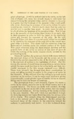
The photograph from a split bicusjDid, Figure 114, shows a
cavity in the mesial, and also one in the distal surface and is of
especial interest. The forms are fairly well outlined, showing
particularly in the one on the left of the picture, that the enamel
rods have not fallen out. Yet the clouding of the dentin reaches
to the pulp chamber. The acid, which has percolated through
tbe decaying enamel, has begun dissolving away the calcium salts
of the dentin. This extends along the dento-enamel junction,
both to the occlusal and to the gingival. In this picture the
backward decay of the enamel, in the extension toward the
occlusal, is particularly well shown. It is this backward decay