Page 153 - My FlipBook
P. 153
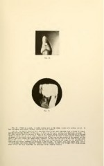
Fig. 78. Caries of enamel. A wnite carious spot on the distal surface of a central
has very sharp and definite outlines, though not very regular.
Fig. 79. An incisor removed for a girl nine years of age, split through areas of decay and show-
ing the broad pulp chamber of that age. A decay in the mesial surface had destroyed the pulp. In
the distal surface there is an area of decay in the enamel, which, superficially, was similar to the one
shown in Figure 78, but not so white. This is very typical of the form of these smooth surface decays
of enamel in its conical shape, with the broad base of the cone on the surface of the enamel and the
apex of the cone toward the dento-enamel junction. In this case the apex of the cone has just pene-
trated the enamel. A little solution of the lime salts of the dentin has begun by the percolation of
acid from the surface through the thickness of the enamel. No enamel rods have fallen away and no
microorganisms have been admitted. Notice that a delicate hyaline zone fringed with shade streaks
away toward the pulp, following the direction of the dentinal tubules.