Page 152 - My FlipBook
P. 152
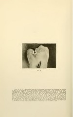
Figs. 75, 76, 77. Photographs from split teeth showing progressively the beginning and progress
of decay of the enamel in pits in the occlusal surfaces of molar teeth. Each illustration is from a
different tooth. Figure 75 represents almost the earliest beginning of caries in the pit, shown by the
whitening of the enamel of the walls of the pit, that can be distinctly recognized in a photograph.
Figure 76 is a more distinctive showing of decay made by the deeper whitening of the enamel about the
pit and the appearance of slight solution of its walls. In Figure 7 7 more decided advance has been
made in the whitening of the enamel and loss of substance in the walls of the pit. The acid has, in
this case, passed the dento-enamel junction and an effect in the dentin is seen. In this tooth there is
also a smooth surface decay of the enamel beginning in the mesial surface, which has also been cut
through centrally. This shows as a faintly whitened area, broad on th? surface and penetrating deepest
in its central part. Its form is characteristic of smooth surface beginnings of decay of the enamel, and
is placed here in sharp contrast with the forms of beginning decay of enamel in pits.